Researchers develop innovative platform for modeling human muscle diseases in worms


Researchers from Bar-Ilan University, in collaboration with Sheba Medical Center, have developed a novel platform to model human muscle diseases in the C. elegans worm. This innovation facilitates the study of diseases in a versatile, scalable way, opening the door to more personalized approaches to disease modeling.
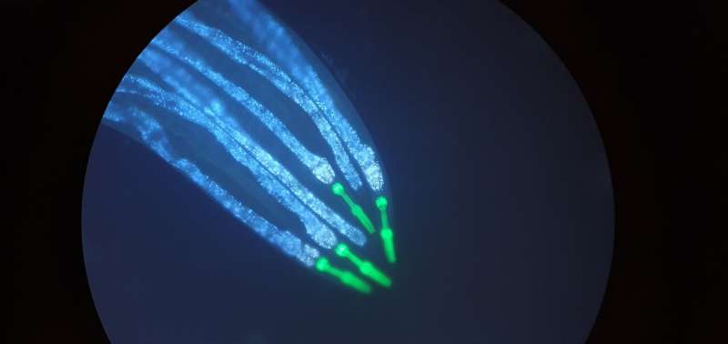
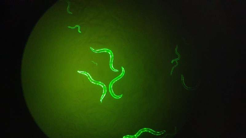
The research team, led by Prof. Chaya Brodie, from Bar-Ilan’s Goodman Faculty of Life Sciences and Institute of Nanotechnology and Advanced Materials (BINA), and Prof. Sivan Korenblit, from Bar-Ilan’s Goodman Faculty, in collaboration with Dr. Amir Dori, a muscular dystrophy specialist from Sheba Medical Center, harvested extracellular vesicles from patients with Duchenne muscular dystrophy and transferred them to C. elegans worms. The results were remarkable, with the worms developing muscle atrophy akin to human symptoms.
This achievement was made possible by a novel approach that utilizes extracellular vesicles derived from patient blood samples. “Unlike traditional methods that rely on genetic modifications in model organisms, this technique relies on elements secreted into the blood,” says doctoral student Rewayd Shalash, who carried out the research with doctoral student Coral Cohen and Dr. Mor Levi-Ferber.
Extracellular vesicles, a form of tiny membrane-enclosed cellular bubbles, contain a rich sample of molecules that represent cellular content. This feature enables the transfer of information across different species, allowing researchers to study human diseases in worm models.

“We transferred the extracellular vesicles and observed the development of muscular dystrophy with significant muscle degeneration, confirming that we discovered a new way to model diseases without the need to alter specific genes,” says Prof. Korenblit.
The study, published in the journal Disease Models & Mechanisms, focused on Duchenne and Becker muscular dystrophy, genetic diseases that affect skeletal muscles, heart muscle, and other muscle groups. The impact of these diseases is profound, leading to disability and premature death. Prof. Brodie says the new platform is expected to facilitate reliable disease modeling and support more effective drug screening. It also has the potential to be applied to other diseases beyond genetic disorders.
More information:
Rewayd Shalash et al, Cross-species modeling of muscular dystrophy in Caenorhabditis elegans using patient-derived extracellular vesicles, Disease Models & Mechanisms (2024). DOI: 10.1242/dmm.050412
Citation:
Researchers develop innovative platform for modeling human muscle diseases in worms (2024, May 15)
retrieved 16 May 2024
from https://medicalxpress.com/news/2024-05-platform-human-muscle-diseases-worms.html
This document is subject to copyright. Apart from any fair dealing for the purpose of private study or research, no
part may be reproduced without the written permission. The content is provided for information purposes only.


